Xiangyu Yin

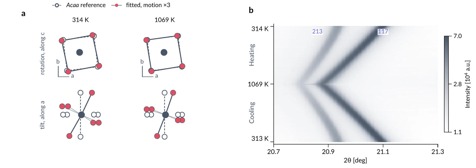
Two diffraction peaks of Ca₃Ti₂O₇ heated to 1069 K and back, and the octahedral rotation and tilt refined from all 346 patterns at once.

Octahedra turning in a perovskite on heating and cooling

Two diffraction peaks of Ca₃Ti₂O₇ heated to 1069 K and back, and the octahedral rotation and tilt refined from all 346 patterns at once.

Yin et al., arXiv:2610.28564, Figure 5 (panels a and b)
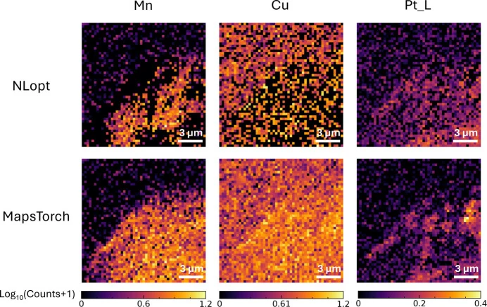
Manganese, copper and platinum maps fitted pixel by pixel from X-ray fluorescence spectra; a standard optimiser above, MapsTorch's gradient-based fit below.

Elemental maps at very low counts

Manganese, copper and platinum maps fitted pixel by pixel from X-ray fluorescence spectra; a standard optimiser above, MapsTorch's gradient-based fit below.

Yin et al., J. Synchrotron Rad. 33, 235–245 (2026), Figure 7

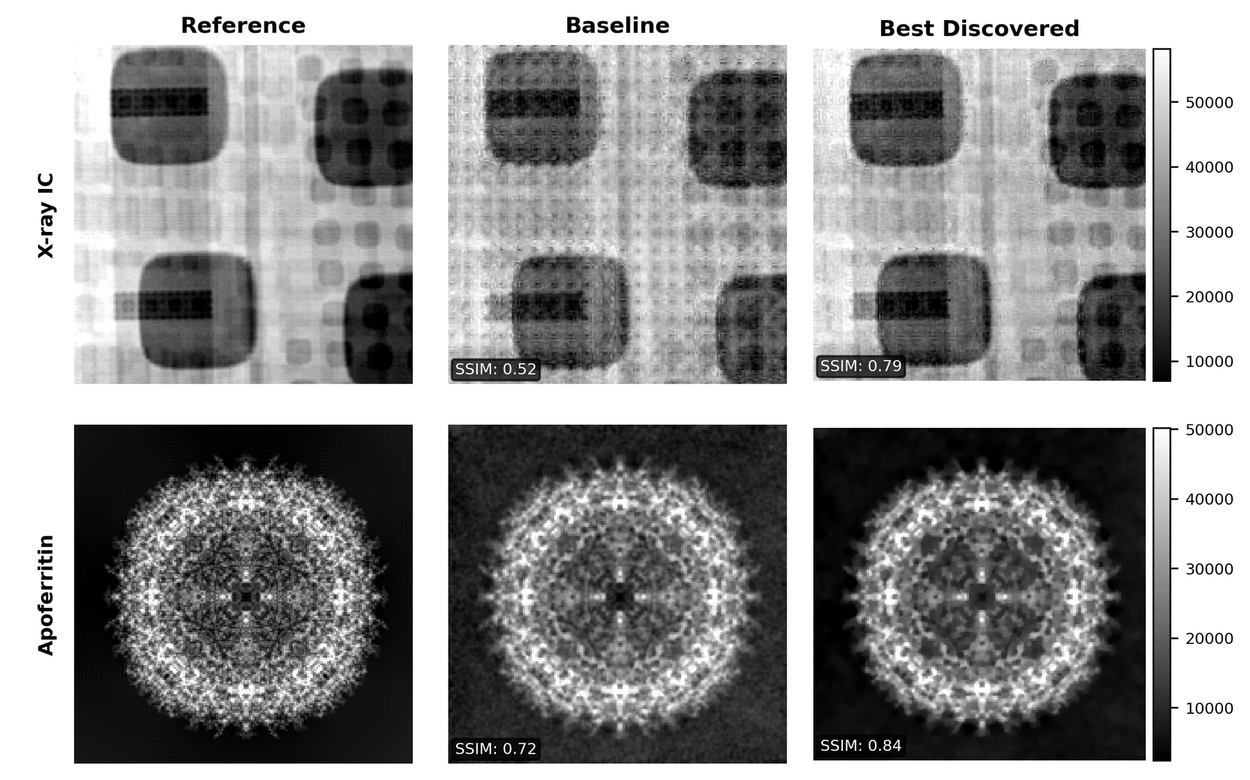
Images recovered from coherent X-ray diffraction by the standard algorithm and by one an LLM-driven search discovered; similarity to the reference rises from 0.52 to 0.79 and 0.72 to 0.84.

Algorithms discovered for ptychography

Images recovered from coherent X-ray diffraction by the standard algorithm and by one an LLM-driven search discovered; similarity to the reference rises from 0.52 to 0.79 and 0.72 to 0.84.

Yin et al., arXiv:2603.05696, Figure 2 (top two rows)
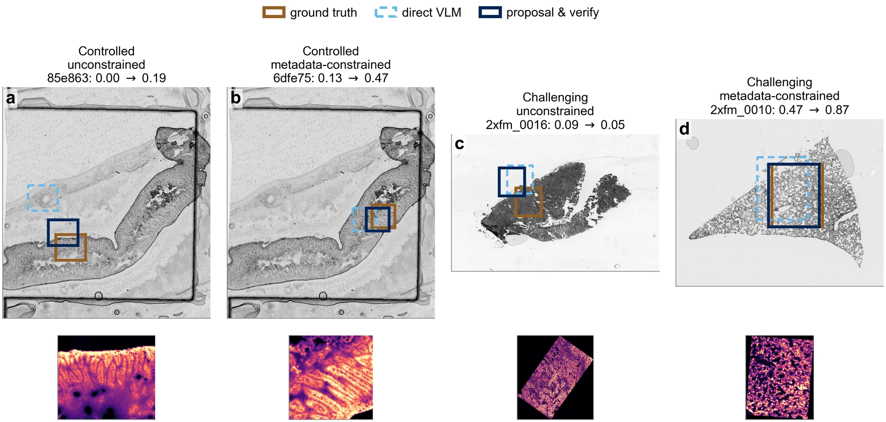
Where on the optical image was each X-ray fluorescence map taken? Boxes from direct prompting of a vision-language model and from an agent that proposes and checks, against the true location.

Finding an X-ray map in an optical image

Where on the optical image was each X-ray fluorescence map taken? Boxes from direct prompting of a vision-language model and from an agent that proposes and checks, against the true location.

Yin et al., arXiv:2608.18309, Figure 3

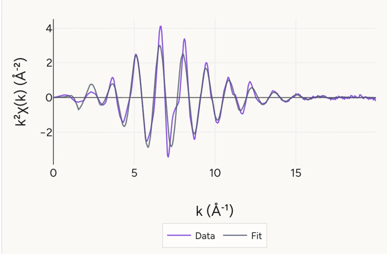
A first-shell fit of metallic cobalt's X-ray absorption oscillations, run by Dr. XAS, an analysis agent in use at the Advanced Photon Source.

Cobalt's absorption fine structure, fitted by an agent

A first-shell fit of metallic cobalt's X-ray absorption oscillations, run by Dr. XAS, an analysis agent in use at the Advanced Photon Source.

Du et al., arXiv:2609.36362, Figure 4 (k-space fit panel)

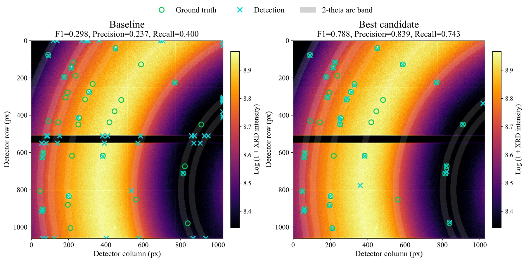
Bragg spots found on one detector frame by the starting detector and by the one an LLM-driven search evolved; the F1 score rises from 0.30 to 0.79.

A search-evolved detector for diffraction hotspots

Bragg spots found on one detector frame by the starting detector and by the one an LLM-driven search evolved; the F1 score rises from 0.30 to 0.79.

Du et al., arXiv:2605.11359, Figure 4